

From Section 6, the recommended diagnostic and monitoring test battery is collated in Fig. 5. Symptoms and at least one positive result of the markers of homeostasis listed below should constitute the diagnosis of DED. If a patient has dry eye symptoms, DED is diagnosed when at least one homeostasis test result is positive. This can occur even if the practitioner does not have access to the full battery of recommended tests. However, if the practitioner has access to only a limited number of the homestasis marker tests and these show negative results, a referral may be necessary to confirm the results of the remaining measures, to which the practitioner does not have access, before a diagnosis of DED can be excluded.
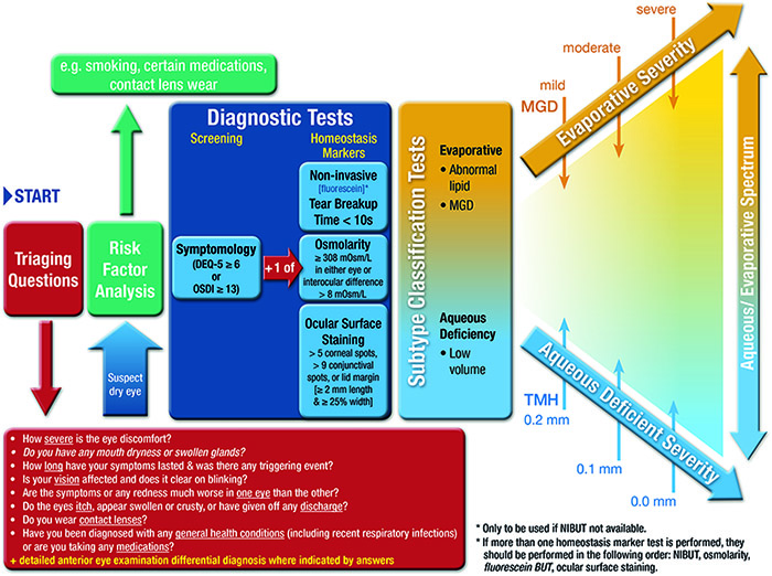
In situations where there are chronic symptoms but limited signs, that are refractory to treatment, then neuropathic pain rather than DED should be considered. Asymptomatic patients with DED type signs, unattributable to other conditions via the differential diagnosis and comorbidities triaging questions in Section 9, might still warrant prophylactic ocular surface treatment. Videos of these diagnostic as well as sub-classification techniques of MGD, lipid thickness/dynamics and tear volume are available on the TFOS website (https://www.theocularsurfacejournal.com).
Tables of severity describing several signs and symptoms and (often arbitrary) cut-offs for different levels are of limited use, as features of dry eye often do not show strong association. Hence it is recommended that severity, for the purpose of selecting treatment, is based on subtype classification features (MGD, lipid thickness/dynamics and non-invasive tear volume) along with symptomology.
The recommended order and clinical practice procedural recommendations are as follows:
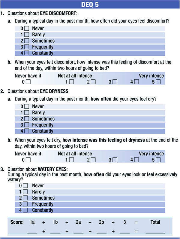
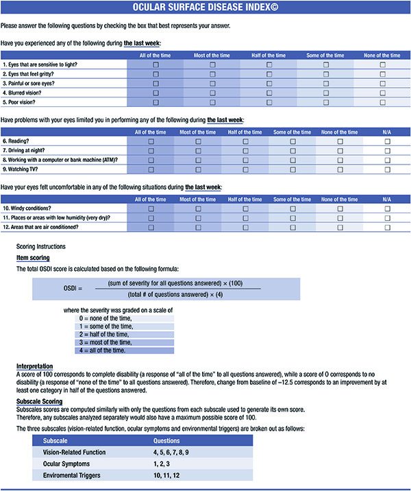
DEQ-5 (Fig. 6a) or OSDI (Fig. 6b) – self-administered [35,37]. Positive result is a DEQ-5 score ≥6 [37], or OSDI score ≥13 [35].
Non-invasive breakup time should be performed with a method where as much of the naturally exposed cornea as possible is specularly illuminated with a light source allowing observation of breakup over the whole surface, after a blink. Objective methods are preferred, with three measurements being performed and the median value recorded. Following training, if a patient can no longer refrain from blinking before the tear film breaks up, this is typically counted as the breakup time for that measurement [194]. The lower median breakup value of the two eyes should be considered in making the diagnosis. The cut-off for a positive finding can be as low as 2.7 s for automated algorithms [142], and up to 10 s for subjective observation techniques [134].
8.2.2 FBUT
FBUT can be considered when non-invasive techniques are not available, but due to its more invasive nature, should follow after osmolarity measurement. Fluorescein should be instilled at the outer canthus to avoid ocular surface damage (see Section 8.4.1 for instillation instructions), with the excess saline on the strip shaken off, or a reduced area fluorescein strip used [118]. For optimal results, viewing should take place between 1 and 3 min after instillation [279]. A positive finding has been reported to be a value < 10 s [13] although in some studies the average in healthy middle aged patients is noted to be lower than this [244].
Osmolarity should be assessed with a temperature stabilised, calibration checked device. In the case of the Tearlab, temperature stability is achieved by having the device powered on for a sufficient period of time with test cards adjacent to the device for at least 30 min. Seat the patient with chin tilted upward and eyes directed toward the ceiling. Place one hand on the face for stabilization, as appropriate. Do not pull the eyelid down or away from the eye. Sample from just above the lower eyelid tear meniscus, being careful not to press inward to avoid contact with the globe during collection. The difference between the eyes as well as the absolute measures can be diagnostic [170,171]. A positive result is considered to be ≥ 308 mOsm/L with the currently available device in either eye [13,15], or an interocular difference >8 mOsm/L [171].
Principally for assessing conjunctival and lid margin damage, a lissamine green strip is wet with saline, with the whole drop retained on the strip for at least 5 s to elute the dye. A 10 μL or ∼ ¼ to ½ of a drop appears to be an optimal volume if pipetting a predetermined concentration solution [272,280]. Otherwise, a drop from the strip is instilled inside the far lower temporal lid in upgaze with the lower eyelid of the eye pulled slightly temporally to avoid damage to the conjunctival or lid wiper tissue (Fig. 7). Studies have suggested that observation should occur between 1 and 4 min post-instillation, and that observation through a red filter potentially aids visualization [272,280]. A positive score is > 9 conjunctival spots [285].
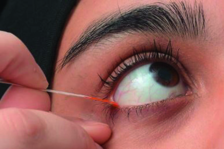
Principally for assessing corneal damage, fluorescein should be instilled in a similar way, but with the excess saline on the strip shaken off to instil a minimal volume. Optimal viewing is between 1 and 3 min after instillation [279]. A positive result is > 5 corneal spots [285].
Lid wiper epitheliopathy can be observed stained with fluorescein, rose bengal or lissamine green dyes, although there seems to be a preference for just lissamine green in recent research, with viewing recommended 3–6 min after repeat instillation using 2 separate strips wet with 2 saline drops [484]. Positive is LWE of ≥ 2 mm in length and/or ≥ 25% sagittal width (excluding Marx's line) [391].
DED severity can change with the time of day so this should be considered in interpreting results and in monitoring DED over time [485,486].